A crushed heel, a smashed knee and a body in which the blood vessels look like a dense mass of plant roots.
On the virtual dissection table, details are revealed with total clarity.
Professor Bengt R Johansson, however, wants to distance himself from anything associated with a pretend reality.
“We are looking at real cases. This is the real thing. This is no illusion,” he says.
Under a black protective cover in a corner of the anatomy room at the Sahlgrenska Academy is a piece of cutting-edge technology. With an accustomed hand,
Bengt R Johansson, professor of anatomy, lifts off the cover.
The virtual dissection table rests on a one-metre high central pillar. It looks like a giant touch screen. And that is what it is – well almost. Infra-red beams are emitted parallel with the screen. By breaking the beams with one or more fingers, Bengt R Johansson changes the image on the screen.
“We used to take dead donors’ bodies and embalm them. I did this myself for over thirty years, the last twenty without the support of any staff. Now the Karolinska Institute medical university in Stockholm performs this function for us, providing 10-15 bodies per year,” says Bengt R Johansson
Recently arrived back in Sweden
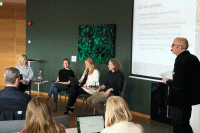
He has just returned from Amsterdam where he attended a meeting with colleagues who also use the virtual dissection table. He would prefer to call it something else.
“We look at images of real cases; images generated by the latest technology. This is the real thing, not something invented or an illusion,” says Bengt R Johansson.
Out in the large anatomy room, the ceiling is high and there is plenty of light. By one of the end walls are rows of refrigerators. Thick doors with heavy metal handles conceal the bodies of the deceased within. Dissection tables, like shallow rectangular stainless steel baths, stand in a line across the floor.
“Our aim is for students to encounter the bodies of the deceased in a sensitive and dignified manner. It is a delicate task,” says Bengt R Johansson.
Crisis reaction
Sometimes students experience a crisis reaction during this encounter.
“Often in these cases a student has recently experienced a loss. His or her grandmother may have died in the preceding months.”
On the whole, however, Bengt R Johansson is pleased at how well students cope when faced with dead bodies even though this is often distressing for modern young people.
“Death is not such a natural presence in our lives these days. At most, only a third of students have ever seen a dead body and this is often because they have worked in the health service before,” he says. He himself grew up on a farm outside Norrköping.
Embalmed bodies
After 15 litres of embalming fluid has been introduced into the arterial system, an embalmed body becomes slightly grey in colour. Clogged blood vessels may cause incomplete embalming of, for example, the lower legs.
The screen of the virtual dissection table is now displaying an image of a foot. The heel has been badly damaged. Bengt R Johansson zooms in by gliding his index finger over the screen. The 3D image we see before us is created from 308 other images.
“What do you think caused that injury?” he asks.
It turns out that the cause was a road traffic accident of the worst kind, a motorcycle crash. Bengt R Johansson points out bone fragments and small, light-coloured, well-defined spots.
“These are air bubbles. Air has entered this foot. This technology is extremely useful for forensic examinations. Strangulation, for example, causes air bubbles and these are normally very difficult to see. Here they show up clearly.”
He brings up an image of a badly injured knee, the result of a riding accident.
The technology could also be used to examine foetuses after spontaneous abortions in order to find the reason for the miscarriage.
3D technology
Bengt R Johansson provides another example of how 3D technology can be used.
“A surgeon would be able to move between the patient and the table during an operation. It is a mobile and user-friendly work station.”
What are its weaknesses?
“Soft tissues do not show up so well. Blood vessels, for example, need a contrast medium.”

He brings up a picture of a body – a skeleton with clearly visible blood vessels. They resemble a plant’s root system with the thicker vessels close to the heart getting thinner as they reach the extremities.
“One thing that is totally necessary is that we create a good image library consisting of real cases. Our primary task is to make anatomy visible but the presence of pathological changes, such as injuries and tumours, has a dramatic and stimulating effect on students,” says Bengt R Johansson.
There are pictures which could already be uploaded both from the Odontology Department and from Sahlgrenska University Hospital. However, the question of personal integrity still remains an obstacle. It is possible, for example, to combine the information in the images to see what the patient looks like.
Not good enough
Another problem is that the data which makes up pictorial medical records is compressed before being stored. These lower resolution images are not good enough.
A virtual light table, a Visualization Table, is produced by the successful company Sectra AB in Linköping. It grew out of research done at Linköping University.
“I myself have not seen the same kind of fruitful collaboration between Sahlgrenska Academy and Chalmers University of Technology. Linköping is light-years ahead of us in this respect,” says Bengt R Johansson.
Hands-on
Despite new technology, even in future students will need to learn anatomy in a more hands-on manner, using the bodies of deceased donors.
“We have a plan, however. We want to combine the actual object to be dissected with images from the same donor’s body on the light table. In order to create these images, we need to add contrast medium to the embalmed body and carry out a full body scan. We are presently discussing the techniques with the Radiology Department,” says Bengt R Johansson.